Posts

Find Cilika’s Latest Breakthrough Product: The Recent Launch Of TF-100 Digital

Image
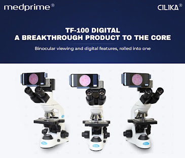
  Unleashing Cilika’sTrinocular Digital microscope from the house of Digital Pioneer MedPrime! The engaging hi-end features of TF-100 smart microscope comprise of upright bright field benchtop Digital microscope with smartphone attachment at trinocular port, simultaneous viewing through Binocular Eyepiece and smartphone display, TrueView technology for capturing 100% circular field of view via smartphone camera. Other general features of microscope includes a Siedentoptrinocular head with Diopteradjustment, Quadruple Nosepiece With High Precision Inner Position Stop Click, Four High Contrast Infinity Corrected Plan Achromat Objective Lenses (4X, 10X, 40X and 100X), Low Position Coaxial Coarse and Fine Focusing System, Safety Rackless Mechanical Stage, Condenser with Iris Diaphragm, Helicoid mechanism, 3W LED Light with Aspheric Collector. Furthermore, TF-100 Trinocular Digital microscope is equipped with user-friendly applications for your smartphone to perform tasks like Data manage

Cilika’s Role & Findings In Preserved Samples | Medprime

Image
  Cilika® Benchtop Series  -  Cilika® Benchtop Series Digital Microscope  comes in three models, available in standards and premium variants. The series is a unique juxtaposition of conventional and digital microscopes with hi-end specifications like 4X, 10X, 40X, and 100X (oil immersion) infinity-corrected high contrast achromat lenses with internal magnification of 10X, interchangeable with binocular head, and premium variants being upgradable for incorporating fluorescence, darkfield, and phase contrast microscopy. The Benchtop series microscopes are ideal for routine use in diagnostic labs and hospitals for testing all kinds of samples like blood, urine, tissues, semen, microbes, smears, etc.,dependent on appropriate ways and means of sample preservation. Moreover, its thoughtful and hi-tech design places the display screen at eye level preventing cervical spondylitis, neck and back pain. Medprime Technologies , started way back in 2014, brain behind Cilika® series of digital micro

How To Preserve Pathology Samples For Longer Duration?

Image
  Generally speaking, collecting and preserving biological samples examined through laboratory microscope and pathology microscope for advanced genetic manipulation and clinical scientific research and molecular study, entail a collection of ultra-low temperature devices for storing and slowing bioprocessing within blood and tissue specimens. Sample collection and DNA preservation are essential for studying specimen’s population and to comprehend biospecimen science, preservation procedure requiring collection, statistical analysis and examination process. Samples Preservation Procedures: Cooling & Refrigeration (Cryogenic & ULT Storage) Drying Desiccants Chemical Preservation Tissue Preservation Liquid Preservation Other Bodily Preservation Cryogenic Sample Preservation Read More at: Digital Microscope

Cilika–Portable Digital Inverted Microscope

Image
  The invention of microscopes nearly a century ago opened up a new branch of science, microbiology. This invention led scientists to observe, examine and work with tiny organisms. So began a new era in healthcare, agriculture, and other related industries, where microscopes are mandatory. For instance, a standard blood test to diagnose blood groups or RBC count prescribed by doctors requires microscopes. The samples collected from the patients at the collection centres are transported to tier-1 cities for microscopic analysis. Transportation is a disadvantage as it increases the chance of sample degradation and cost and delays the treatment.  Microscopes play a vital role in education as well. Most of the colleges and private education sector can procure binocular microscopes. Higher-end, high-cost research-grade microscopes with advanced features are not feasible for all educational institutions. Considering that a practical laboratory session is very crowded, time is limited, and mi

Why Digital Microscopy?

Image
A digital microscope is a modern microscope that lacks an eyepiece, an immense difference from the traditional optical microscope. They have a digital camera which is the detector and the image output device. The display of the image is done on a computer screen or monitor, defining the microscope’s digital scope. This microscope’s light source is an inbuilt LED-source as compared to the optical microscope whose light source is accessed from outside the microscope through an eyepiece. Therefore, in the digital microscope, human optics access is eliminated since the entire apparatus has an image monitoring system. There are variants of the digital Microscope including the USB digitalized microscopes which are very expensively industrialized digital microscopes, such as the Kohler illumination and the Phase-contrast Illumination, with installed webcams and macro lens. The first Digital Microscope was manufactured in 1986 in Tokyo, Japan which constituted a control box and a lens the was

What are the future telemedicine trends to look out for in India?

Image
  Telemedicine is also referred to as “telehealth” or “e-health”. Rapid advancements in connected care technology, big data and analytics are continuing to move telemedicine into the mainstream as it allows healthcare professionals to evaluate, diagnose and treat patients in remote locations using telecommunications technology. The approach towards this has evolved over the last decade and its adoption rate has significantly improved. Covid-19 has certainly changed the telemedicine future trends! Though traditionally telemedicine is viewed as a tool to bridge the accessibility gap to provide care to remote areas, the capabilities and full potential of telemedicine are yet to be realized. A patient-centric way to provide quality healthcare services, telemedicine has yielded an opportunity for standardization and equity in the provision of healthcare. As India is striving to achieve sustainable development goals, using all the available resources is important to be able to provide univer

WHY UPGRADE TO A DIGITAL MICROSCOPE?

Image
Expert Consultations: Digital microscopy makes it much easier to obtain second opinions or expert consultations for unclear cases via the sharing of images and videos. Evidence-based Reporting: More and more labs are moving towards evidence-based reporting, with images of the sample attached along with the patient’s report. Online Discussions and Classes: It allows seamless live discussions/seminars with colleagues or students via screen sharing, particularly now that offline classes and conferences are not possible. Virtual library: Digital microscopes facilitate capturing images of unique and special cases for creating libraries for educational purposes. Health Benefits: Viewing samples on a screen as opposed through binoculars for hours continuously will help avoid cervical spondylitis and unwanted stress on the neck, back, and eyes.  Read More At:  Digital microscopes